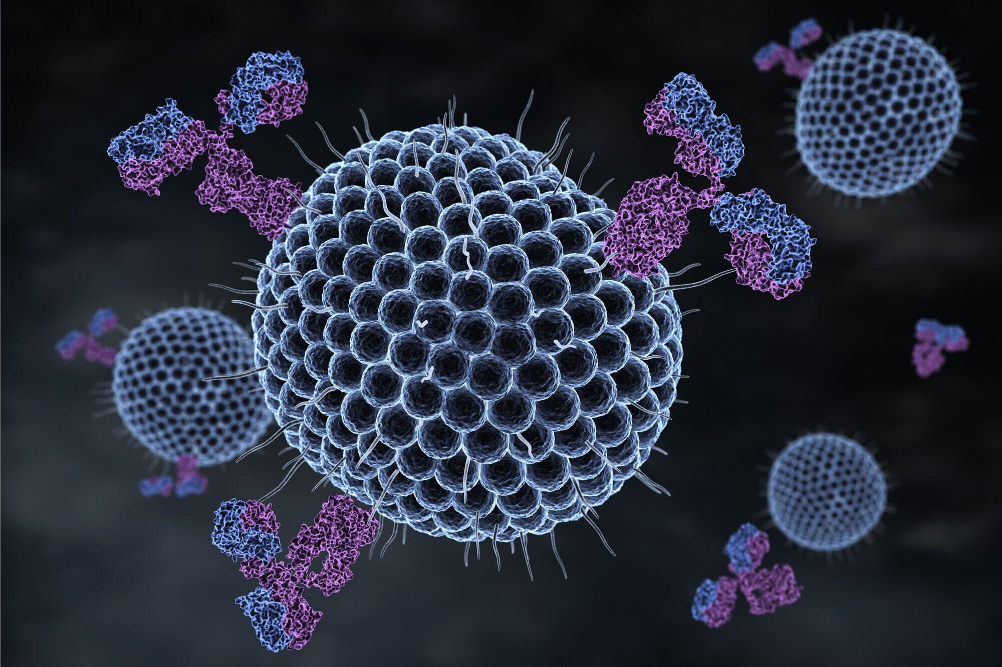
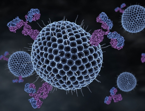
This powerful technique enables rapid identification and sorting of cells, streamlining the manufacture of many biologic pharmaceuticals.
by Raymond E Peck, CEO of VxP Biologics
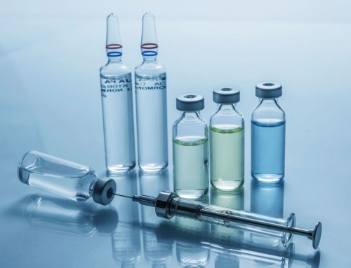
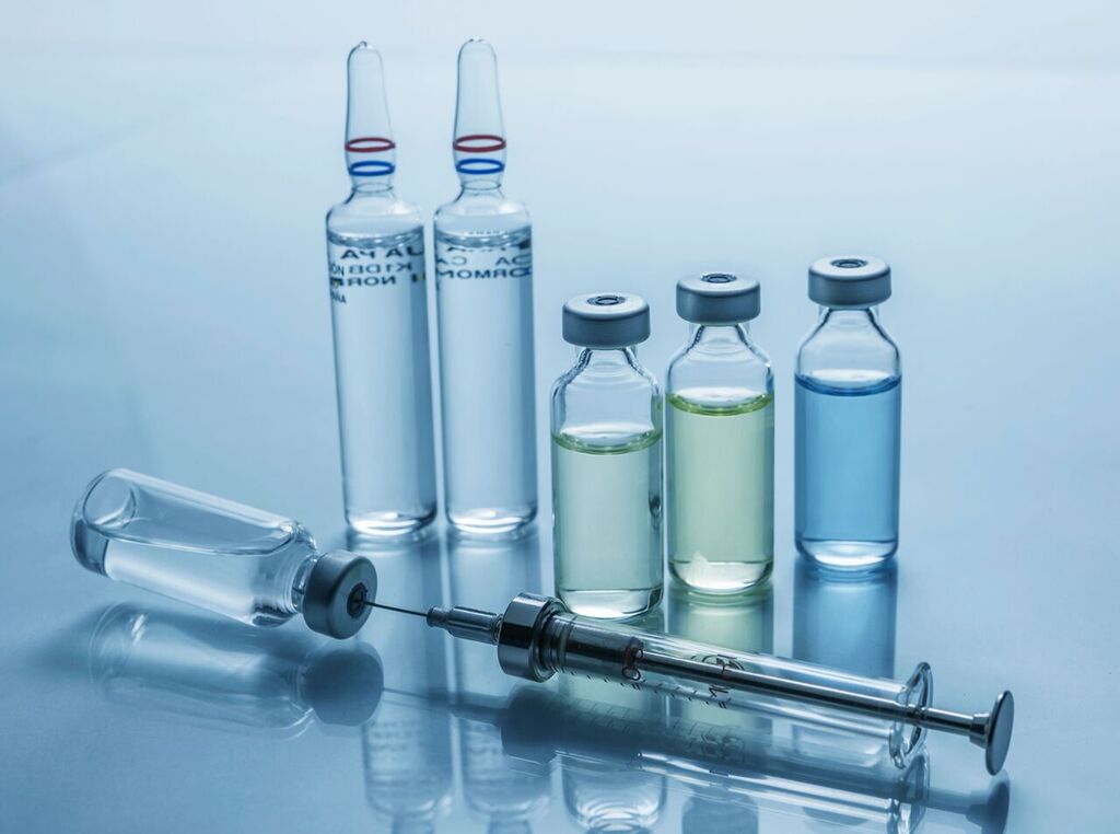
In the manufacture of many types of biologics, flow cytometry is a crucial tool for identifying, analyzing and sorting cells. This technique can provide a wide range of data points about cells’ physical and chemical attributes, including their volume, cytoplasmic granule content, and even nuclear structure.
The underlying principle is to label cell components with fluorescent dye (typically carboxyfluorescein succinimidyl ester, or CFSE), then use a hydrodynamically focused stream of fluid to shuffle those cells in single file across the beam of a laser. The laser excites fluorescence in the dye, emitting light at a longer wavelength than that of the source. By emitting laser light at varying wavelengths, the cytometer sends a combination of scattered and fluorescent light back to its detectors, providing data which is then analyzed by computer software to provide information about many properties of each cell.
As complex as the procedure is, the detector and software are powerful enough to perform it thousands of times per second. With a combination of Forward Scatter (FSC) detectors in front of the stream, and Side Scatter (SSC) detectors perpendicular to it, the cytometer enables engineers not only to classify thousands of individual cells in a matter of seconds, but also to track their proliferation, apoptosis, and other cell cycle properties.
Flow cytometry can aid in all the following applications within a biologic manufacturing pipeline.
A flow cytometer can help identify, quantify, and sort thousands of cells per second.

Flow cytometry can be used to track cell proliferation and interaction.
The manufacture of biologic products often involves the generation of a variety of cells, including blood, bone, marrow and lymph; but not all these cell types will be desirable in the final product. The process of identifying and quantifying cell populations (so they can be sorted) is known as immunophenotyping.
This process is one of the most widespread uses of a flow cytometer. Proteins specific to each cell population are labeled using a fluorescent tag that adheres to the cell’s outer membrane. The flow cytometer then causes cells with these proteins to send fluorescent light to its detectors, enabling these cells to be filtered out.
Many flow cytometers also include built-in cell sorters, which can physically separate target populations of cells, and isolate them in collection vessels such as test tubes. As the cytometer analyzes each cell that passes single-file through its laser beam, the sorter automatically “kicks” all cells of interest out of the stream and into the tube, thousands of times per second.
Flow cytometry can also aid in analysis of cell cycles and death conditions.

Flow cytometry can also aid in analysis of cell cycles and death conditions.
Cells undergo internal and external structural changes throughout all four phases of the cell cycle. By labeling cell components with fluorescent dye, then running the cells through a flow cytometer, engineers can track the replication states of entire cell populations, determining statistical distributions of each phase of the cell cycle, and even providing early-warning detection of chromosomal abnormalities that may lead to issues in the manufacturing pipeline.
Eukaryotic cells may die due to apoptosis, in which they initiate their own internally programmed death; or due to necrosis, an abnormal condition brought on by physical and/or chemical damage. The successful manufacture of a biologic product requires the ability to precisely and reliably distinguish between these two states. Flow cytometry can track abnormal changes in cell morphology, biochemistry and/or molecular composition, all of which may indicate necrosis. By providing clear statistical data on proportions of cells undergoing apoptosis and necrosis, a flow cytometer can enable researchers to identify causes of the latter condition, and act to address them.
Flow cytometry can even be used to track cell proliferation and interaction.
In a biologic manufacturing pipeline, cell proliferation assays are useful for measuring cells’ metabolic responses to growth factors, cytokines, media components and other potential growth factors. A flow cytometer is highly useful for measuring cell proliferation. Engineers first label cell membranes with fluorescent dye, then run the cells through the cytometer as they begin to undergo mitosis, passing half the dye to each of their daughter cells. The diffusion in fluorescence provides a precise indicator of rates of cell activation and proliferation.
At certain stages of the manufacturing pipeline, it may also be useful to track the degree to which various populations of cells respond to an external stimulus. When such a response occurs, membrane-bound ion channels on the cell membrane pump calcium into the cell, causing a spike in the concentration of intracellular calcium, which delivers energy for a response to the stimulus. Thus, by monitoring the flow of calcium into each cell, a flow cytometer can rapidly assess the responsiveness of cell populations to a stimulus that has been introduced.
All these applications of flow cytometry demonstrate how effectively the technique enables the rapid identification, sorting and analysis of cells; as well as diagnosis of common problems with cell proliferation, responsiveness and death. By providing clear statistics on cell structure and phase, and reporting any abnormalities, flow cytometry helps detect, address, and even prevent many issues in a biologic manufacturing pipeline.
